Internal Anatomy of the Crayfish

![]()
Directions: Follow the directions step-by-step, locating each of the structures in the order they appear in the directions. You may want to review or reference terms you don't remember (It is expected that you know the terms: cephalothorax, thorax, rostrum, and dorsal before you start this dissection). You may use your book diagrams to help you locate the organs. *Check the box next to the number when you have completed that step.
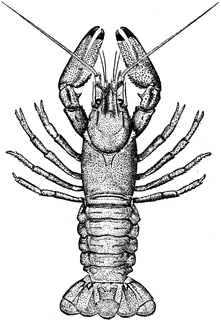
1. Place the specimen in the dissecting tray dorsal side up.![]()
2. Carefully insert the point of the scissors under
the top of the carapace (shell) at the back of the cephalothorax and cut
up the middle to the rostrum. Peel slowly to preserve the organs lying underneath the carapace. ![]()
3. Cut acrss the carapace just back behind the eyes and
remove the two pieces of the carapace. ![]()
4. Note the exposed gills. (Feathery like structures just under the carapace you just remove.) ![]()
5. Remove the exposed gills and legs attached to the thorax. Carefully
separate the dorsal layer of muscles in the thorax and note the light
colored heart just underneath. ![]()
6. Remove the heart. The two light colored masses extending
on each side of the body into the head are the digestive glands.
(The heart is located just posterior to these.) ![]()
7. Between the digestive glands, you will find the small
pair of white reproductive organs and cuts in the male animal. If your
specimen is female, you will probably see a large mass of dark colored
eggs. ![]()
8. To locate the intestine, insert the point of the scissors under the dorsal side of the shell of the abdomen and cut back to the telson. Spread the shell, and the intestine will be found as a tube on the tops side of the muscles of the abdomen. ![]()
9. Trace it forward to the point where the intestine joins the large, thin walled stomach in the front part of the cephalothorax. ![]()
10. Now, remove all the organs in the thorax by cutting
the short esophagus below the stomach and the bands of muscle holding
the stomach just back of the eyes. You should be able to lift out most
of the internal organs in one piece. ![]()
11. Clean out the remaining tissue in the head so that the green glands (kidneys) are exposed. ![]()
12. In the front part of the head cavity, between the eyes,
not the small mass of white tissue, the brain. ![]()
13. Trace the nerves that go from the brain to the antennae
and eyes. ![]()
14. Cut the hard tissue on the flore of the thorax with
your scalpel so that you can follow the ventral nerve cord back
from the brain to the abdomen. ![]()
15. Spread the shell of the abdomen apart and pull out the
large muscle. (This is the part of the body eatern in shrimp and lobsters.) ![]()
16. Note the nerve cord that is now exposed on the floor
of the abdomen. The enlargements of the nerve cord in each segment of
the abdomen are the ganglia. ![]()
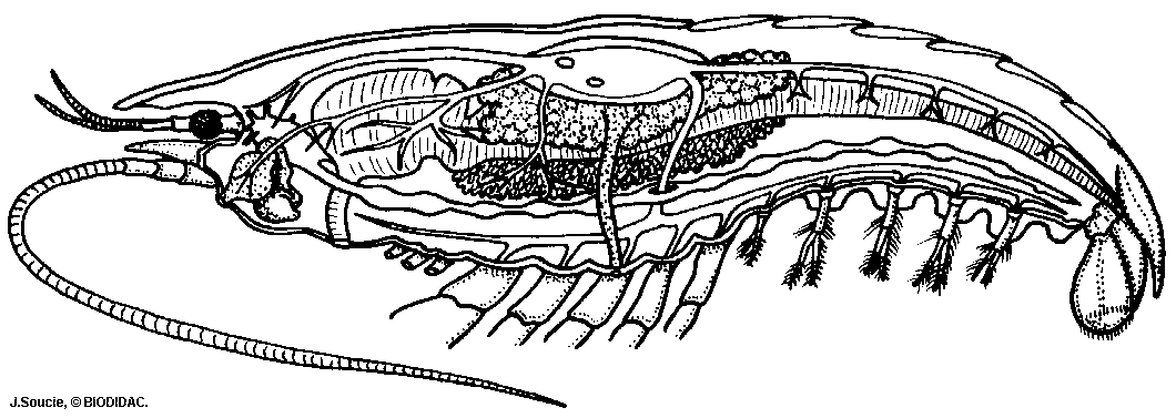
Label as many structures as you can on the picture.
Other Resources on Arthropods
Arthropod Coloring - color the body parts of different kinds of arthropods
Crayfish External Anatomy – focusing on the appendages and mouthparts
Crayfish Virtual Dissection – images and walk-through of crayfish dissection
Grasshopper Anatomy – examines the appendages and mouth parts