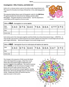
In this activity, students use a codon chart to compare the DNA sequence of HbA (normal hemoglobin) to HbS , the hemoglobin found in blood of sickle cell patients. The DNA differs in a single base, where the codon for normal hemoglobin codes for glutamine, and the mutant form codes for valine.
The worksheet is fairly basic, intended to help students understand the relationship between DNA, RNA, and proteins, “The Central Dogma.” A codon wheel is included on the chart but could be replaced with a codon chart. The concept of mutations is included, but it is not the focus of the activity. I have a similar, more advanced activity, that does focus on mutations: Investigation: DNA, Proteins, and Mutations.
A final question in the synthesis section is specific to NGSS standard: Construct an explanation based on evidence for how the structure of DNA determines the structure of proteins which carry out the essential functions of life through systems of specialized cells. ( HS-LS1-1 ). Students must fill in blanks to answer the question, though you could make it more challenging by not providing the blanks.
This activity can be paired with HHMI’s “The Making of the Fittest: Natural Selection in Humans” which explores how the sickle cell trait provides resistance to malaria and why this gene persists within the population.
Grade Level: 9-12
Time Required: 15-20 minutes
HS-LS1-1 Construct an explanation based on evidence for how the structure of DNA determines the structure of proteins which carry out the essential functions of life through systems of specialized cells

