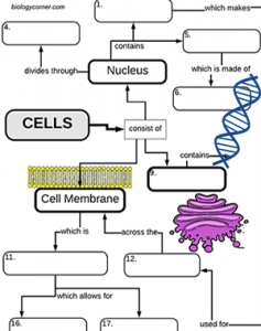
This graphic organizer (concept map) organizes the cell structures around the three main parts of the eukaryotic cell: the nucleus, cytoplasm, and cell membrane. Structures and their functions are included, such as the golgi apparatus and the mitochondria.
Processes, such as diffusion are considered in the area near the cell membrane. A word bank is included, but you could remove it to make the exercise more challenging.
Graphic organizers are very helpful in studying biology and anatomy, however, it provides greater benefit when students make their own. I start off the year with pre-made graphic organizers like this one or the one on the body systems, then transition students into being able to create their own.
For example, once the anatomy students got the chapter on tissues, they have already seen a few of these graphics and are now tasked to make their own on the tissues types of the body.
From a pedagological point of view, students creating their own concept map is probably more effective than the fill-in version. However, students need to practice and model the process before they are ready to create their own.
HS-LS1-2 Develop and use a model to illustrate the hierarchical organization of interacting systems that provide specific functions within multicellular organisms

