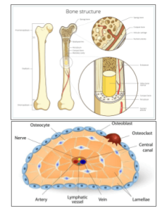
To fully understand how bones function, it’s essential to look at the structure of a long bone. Long bones, like the femur or humerus support the body’s weight and facilitate movement. They have several distinct layers and regions, each with a specific role. Within the bones, microscopic cells called osteocytes create the rings and the structure of the bone which. Anatomy students learn about bones of the skeleton, often naming each, as well as learning about the microscopic structure of bone cells and viewing bone with a microscope.
Key Structures of the Long Bone
Periosteum: tough, fibrous membrane that covers the outer surface of the bone. It also serves as an attachment point for tendons and ligaments.
- Compact bone: dense, hard layer provides the majority of the bone’s strength and protection. It is composed of tightly packed osteons, which are the structural units of compact bone.
- Spongy bone – lies within the epiphysis, is porous like a sponge with spaces filled with bone marrow, which is responsible for producing blood cells.
- Medullary cavity: runs through the center of the bone shaft (diaphysis) and houses yellow bone marrow, which primarily stores fat.
- Endosteum: thin membrane that lines the medullary cavity and covers the internal surface of the bone.
Key Structures in the Bone Matrix:
The bone matrix is one of the most fascinating aspects of the human skeleton. Furthermore, it is a dynamic, living tissue that provides strength and support, but also flexibility. For students, learning about the bone matrix can daunting, because so many of the words are unfamiliar. The bone matrix is the non-cellular component of bone tissue, where all the key structural elements reside. Think of it like a scaffold that allows bone cells to function and thrive.
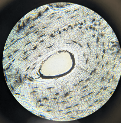
- Osteocytes: These are mature bone cells that sit in small spaces called lacunae within the bone matrix. They help maintain the tissue and communicate with other cells via tiny canals called canaliculi.
- Lamellae: Bone is layered in concentric rings, forming lamellae, which make up the structural units called osteons or Haversian systems.
- Haversian Canal: At the center of each osteon is a canal that contains blood vessels and nerves, ensuring bone tissue gets nutrients and can heal when damaged.
Resources for Learning Bone Anatomy
Bone Matrix Coloring – color the osteocytes, lacuna, spongy bone, and other structures
Color the Long bone – identify and color structures like the epiphysis, diaphysis, and medullary cavity
Label the Structures of the Long Bone – a detailed graphic showing structures with a word bank
Google Slides and Student Notes – includes bone anatomy, skeleton labeling, and disorders with printable sheet for students
View the Bone Matrix with the microscope – prepared slides show osteocytes, canaliculi, and the Haversian canal