
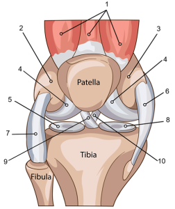
The knee is a complex joint that allows us to walk, run, jump, and move with ease. It connects the thigh bone (femur) to the shin bone (tibia). Ligaments connect the bones are are essential for stability of the joint. In fact, common injuries to the knee occur in sports, such as a tear to the anterior cruciate ligament (ACL).
In this exercise, students identify the major structures of the knee joint, including the cartilage and ligaments. By coloring the location of these major structures, students gain a better understanding of the knee joint.
The coloring activity has each of the structures labeled with letters. Students locate the structures and color according to the directions. For example, students will color the lateral meniscus purple and the medial meniscus pink. This also helps students with directional terms such as medial and lateral.
The second page reinforces learning where students label each of the structures. They can refer back to the previous page to help with the labeling. You can also show students the structures on a model of the knee.
Download the Google doc file to share with your students. I also have the answer key available on TpT.
You can pair this activity with other coloring worksheets on the skeletal system:
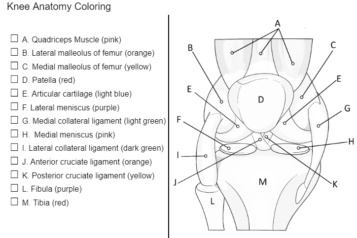
Summary of the Ligaments:
Anterior Cruciate Ligament (ACL) – letter J
- Location: Inside the knee joint, connecting the femur to the tibia.
- Function: Prevents the tibia from sliding too far forward relative to the femur and provides rotational stability. It is one of the most commonly injured ligaments, especially in athletes.
Posterior Cruciate Ligament (PCL) – letter K
- Location: Also within the knee joint, it runs alongside the ACL but in the opposite direction, connecting the femur to the tibia.
- Function: Prevents the tibia from sliding too far backward relative to the femur. It is stronger than the ACL and is less frequently injured.
Medial Collateral Ligament (MCL) – letter G
- Location: Along the inner (medial) side of the knee, connecting the femur to the tibia.
- Function: Provides stability to the inner knee, preventing it from bending too far inward. It helps resist forces that push the knee medially (toward the body).
Lateral Collateral Ligament (LCL) – letter I
- Location: Along the outer (lateral) side of the knee, connecting the femur to the fibula (a smaller bone next to the tibia).
- Function: Provides stability to the outer knee, preventing it from bending too far outward. It helps resist forces that push the knee laterally (away from the body).