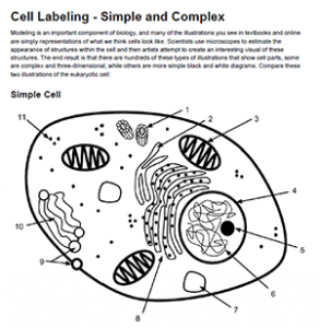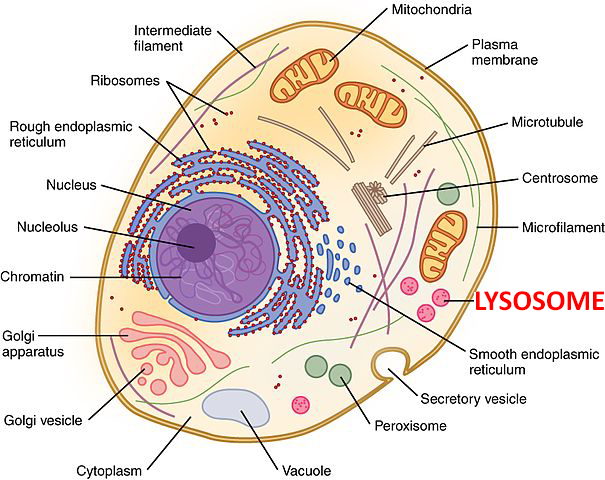
Students practice labeling organelles on a simple model (2D) and a more complex model. The idea is for students to gain an appreciation for how cell diagrams are created. They don’t all look alike, and are often artistically created.
Cell organelles tend to follow basic design rules, like the mitochondria will generally look like a peanut with an internal membrane.
I created this worksheet for cell structure practice. The diagrams are simple enough for even beginner biology students to figure out. You can also put the images on an overhead and help students work out where each of the cell structures are located.

For a more challenging activity, ask students to annotate each structure with its function

