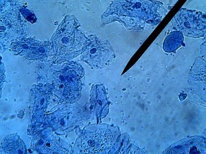
Plant cells are easy compared to animal cells, they don’t move and are arranged in nice blocks. Students can easily find the cells and the nuclei and chloroplasts. Viewing animal cells can be a little bit more challenging.
Fortunately, you have a ready source of animal cells in your students – that is, inside of their mouths. The technique is simple and included in this lab handout, or you can post instructions on the board.
Instructions
1. Place a drop of methylene blue on a slide – this is to stain the otherwise clear cells.
2. Take a toothpick and rub the inside of your mouth. You do not need to press hard.
3. Swirl the toothpick in the blue stain and add a cover slip.
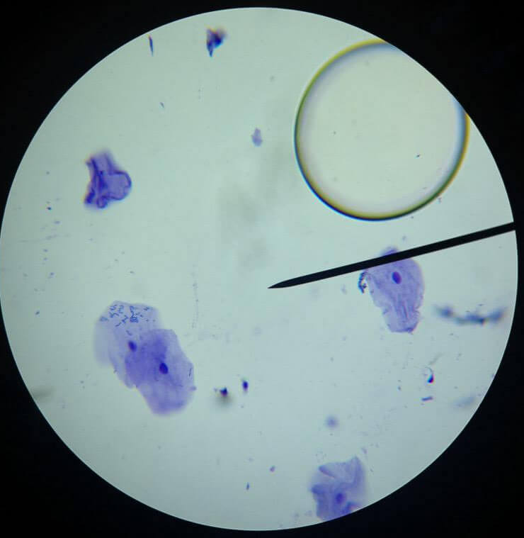
Getting a clear view using the microscope is not as simple. Debris or dust on the slide will pick up the stain and can distract students from finding cells. Air bubbles are also easy distractions, because they are round and about the size of a cell. The image below shows an air bubble in the top right area, the purple blobs are the cells.
If your students have trouble with finding their cells, it may be a good idea to set up a few “stations” with the microscopes already focused on the cells so that students can get an idea of what they are looking for. If you have the technology, you can project the microscope images on a projector. Remind students that cheek cells will not be perfect circles, their shapes are “roundish” but if it looks like a perfect circle, it is probably an air bubble.
Students are often fascinated by the dark blue debris stains which can be dust on the slide, or random stuff in their mouth. You’ll get a lot of “ews” over that.

Students post their “#cellfies” to social media!

