Cat Vessels - Arteries and Veins
During this lab, students carefully teased the muscles and tissues away to reveal the arteries and veins. The goal was to identify each of the vessels on theirlab guide and pass a lab test at the end of the activity where they are asked to locate the vessels.
Cat Vessel Images can be found in this GALLERY
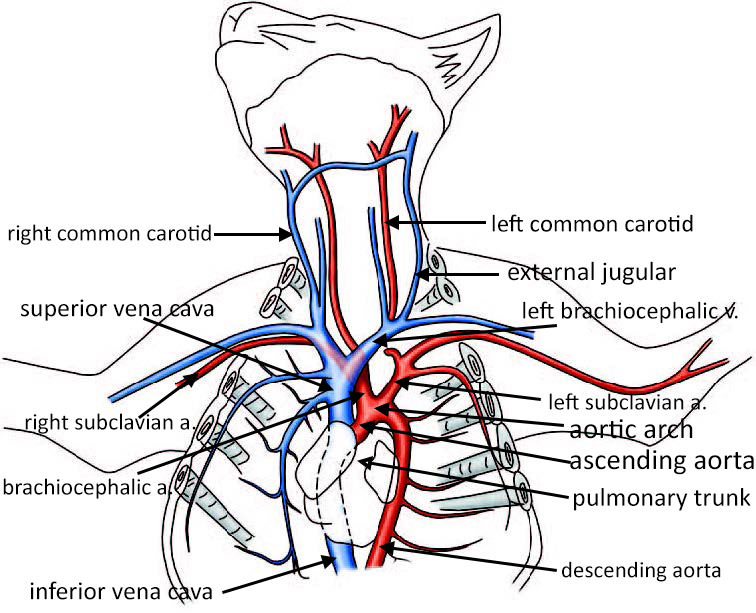
After a Y incision is made, students carefully remove the pericardium and tissue surrounding the heart so that the major vessels can be easily seen: the arch of the aorta, the brachiocephalic artery, the subclavian arteries, and the carotids. On the image below the arteries are colored pink. The veins visible at the top of the heart include the superior vena cava, the brachiocephalic veins (2) and the jugular.

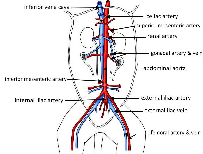
Once the aorta has been revealed, students follow it down down into the abdominal cavity. The inferior vena cava lies next to the aorta and can be identified by its blue color in injected cats. There are three arteries of interest that branch from the abdominal aorta: the celiac trunk, the superior mesenteric, and the inferior mesenteric. Students do not remove the intestines at this point, but instead carefully tease away the tissue with minimal damage to other structures.
Trace the Aorta
Follow the aorta to the lower body to reveal other arteries attached to it. Most of these arteries have veins running parallel.
The kidney sits retroperioneally (or behind the peritoneum), students remove the membrane that surrounds the kidney in order to more easily see the renal artery and vein. The renal vein is often easier to locate, the artery is found next to it.
Students continue to trace the abdominal aorta into the lower abdominal cavity. Careful teasing of the tissue will reveal the internal and external iliac arteries. The iliac vein is located in this region also.
The external iliac can be traced into the leg where it continues as the femoral artery. The femoral vein lies next to it.
