SKELETAL SYSTEM
FUNCTIONS -
support and protection
- body movement - muscles "pull" on bones
- blood cell formation - occurs in red bone marrow
- storage of inorganic materials - calcium, magnesium,
sodium, potassium...
Organization of the Skeleton
- normally 206 bones with 2 Main Divisions: axial and appendicular
1. Axial: head, neck, trunk, vertebrae, ribs, and sternum
2. Appendicular: limbs and bones
connecting the limbs to the:
- pectoral girdle - (scapula & clavicle), upper limbs (arms)
- pelvic girdle - (coxal bones), lower limbs (legs)
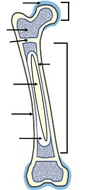 Long Bone Structure:
Epiphysis - ends of bone, forms a joint with another
bone.(1)
Spongy bone - red marrow (3)
Diaphysis - shaft of bone (6)
Articular cartilage- padding covering the ends of bones (2)
Epiphyseal line - growth plate (4)
Periosteum - tough membrane covering over entire bone. (8)
Endosteum - lines interior (3)
Medulla- within diaphysis, contains yellow bone marrow (7)
Label the Bone Structures (Long Bone)
Endosteum- lining of the medullary cavity
- Red Marrow = produces blood cells | Yellow Marrow =fat storage
Two Types of Bone Tissue
1. Compact bone - wall
of the diaphysis, solid, strong
2. Spongy(cancellous) bone - epiphysis.
Microscopic Structure
*Bone tissue is called osseous tissue. - The matrix is composed of collagen and
inorganic salts
- Osteocytes (mature bone cells)
- enclosed in tiny chambers called lacunae
- lacuna form concentric rings called lamella
- Canaliculi (canaliculus)
connect the osteocytes
-
Haversian and Volkmann canals provide passageways for blood vessels
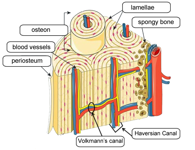
Bone Matrix Coloring (assignment)
Bone Development and Growth:
Bones first form as hyaline cartilage. The cartilage then gradually changes into bone tissue - a process called ossification.
primary ossification center - diaphysis, increase diameter
secondary ossification center - epiphysis, increase length
epiphyseal disk ( growth plate) between the diaphysis and the epiphysis.
Osteoblasts - produce bone cells called OSTEOCYTES
Osteoclasts - dissolve bone tissue - a process called RESORPTION.
Basic Types of Joints (articulations):
1. Synarthrotic - immoveable joint, these junctions are called sututes
2. Amphiarthrotic- slightly moveable joint, vertebrae
3. Diarthrotic - freely moveable jointBall & Socket (shoulder, hip)
Hinge (elbow, knee)
Pivot (lower arm)
Saddle (thumb)
Bones of the Skull
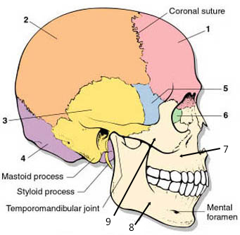
1. Frontal - anterior portion (forehead)
2. Parietal - one on each side of the top part of skull
3. Temporal - side, above ear
4. Occipital- forms the back of the skull
5. Sphenoid - within the cranium, partly visible in eye socket
6. Ethmoid - nasal cavity, visible in eye socket
7. Maxilla - forms upper jaws
8. Mandible - lower jaws, only moveable bone of the skull
9. Zygomatic - cheek bones
10. Vomer bone - divides nasal cavity
11. Palatine bone - roof of the mouth
12. Ethmoid bone - roof of nasal cavity
Sutures - connection points between skull bones
1. Coronal - between frontal and parietal bones
2. Lambdoidal - between occipital and parietal bones
3. Squamosal - between temporal and parietal bones
4. Sagittal - between parietal bones
Fontanels - "soft spots"
of an infant's skull, these form sutures as you age, top spot is the anterior fontanel
Foramen Magnum - Large opening through bottom of skull, where the spinal cord enters skull
The Rest of the Bones
Vertebrae - Cervical (C1-C7) = neck | Thoracic (T1-T12) = middle back | Lumbar (L1-L5) = lower back
Sacrum and Coccyx (tailbone)
Ribs - Thoracic Cage, 12 pairs
Pectoral Girdle: Shoulder. Two clavicles (collar bones) and two scapulae (shoulder blades)
Upper arm - humerus.
Lower arm - radius and ulna
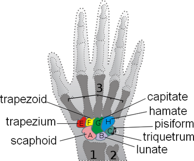
Wrist - 8 small bones called carpals
Hand: Metacarpals | Fingers: phalanges
Pelvic Girdle: Hips. Two large bones called coxae (coxal bones)
Upper leg - femus
Kneecap: patella
Lower leg - tiba and fibula
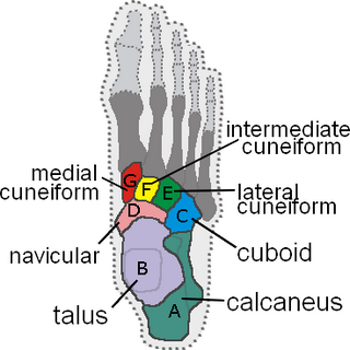
Ankle and Upper foot - 7 bones called tarsals
Largest is the heel bone called the calcaneus
Foot: metatarsals | Toes: phalanges
Label the Bones of the Skeleton
Carpals and Tarsals
Broken Bones
Closed | Open (compound) | Multiple | Comminuted | Greenstick | Spiral