Mitosis in Real Cells
Introduction:
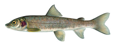
To study mitosis, biologists often look at particular cells. Remember, that mitosis occurs only in areas of growth, so finding a good spot to study it can be challenging. Two specimens are commonly used by biologists to study mitosis: the blastula of a whitefish and the root tip of an onion.
The whitefish embryo is a good place to look at mitosis because these cells are rapidly dividing as the fish embryo is growing. The onion root is also a good place because this is the area where the plant is growing. Remember that when cells divide, each new cell needs an exact copy of the DNA in the parent cell. This is why mitosis is only visible in cells that are dividing, like the whitefish embryo and the onion root tip.
Mitosis can take several hours to complete. Scientists will make slides of cells that should be undergoing mitosis in order to find a particular cell in any of the stages - prophase, metaphase, anaphase, telophase. Remember that most cells you see will be in interphase, that's the cells "resting" state. Your task is to look at photographs of actual slides and identify the stages of mitosis. Answer the questions on each page in your lab notebook or print this first page and answer direction on it.
Tasks:
View slide images of a whitefish blastula and an onion root to see cells in various stages of mitosis. Answer the questions as you read the introduction and view the slides.
You may want to print this page out to answer the questions on it, or just answer questions (#1-5) on your own paper.
Introduction:
1. Why is the whitefish used to study mitosis?
2. What are the four stages of mitosis?
3. How long does it take for mitosis to complete? Why will most of the cells you view be in interphase?
View Cells
Click to View Whitefish Embryo 
Click to View Onion Root Tip 
*Or view real slides showing mitosis - Set from Amazon
4. For each specimen list sketch and label the phases of mitosis
View 1 |
View 2 |
View 3 |
View 4 |
View 5 |
|
| Whitefish | |||||
| Onion |
