Tag: coloring
-
Understanding Knee Anatomy with Coloring
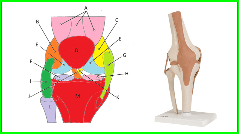
The knee is a complex joint that allows us to walk, run, jump, and move with ease. It connects the thigh bone (femur) to the shin bone (tibia). Ligaments connect the bones are are essential for stability of the joint. In fact, common injuries to the knee occur in sports, such as a tear to…
-

My Coloring Book – Animals of North America
A coloring book showcasing the animals of North America. Shows a variety of animals in their natural habitats!
-
Comparing Monocots and Dicots with Coloring
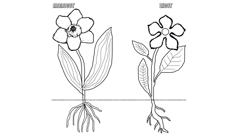
Students learn about monocots and dicots by coloring diagrams of germination and flower structure. Great to pair with germination experiments!
-
Brain Anatomy (Coloring)
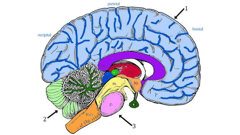
Students learn brain structures and their functions by reading short descriptions and coloring an image. The description are organized into three main areas of the brain: cerebrum, cerebellum, and brain stem. The brain stem also includes structures of the diencephalon (thalamus and hypothalamus). In addition to coloring the image according to the directions, students identify…
-
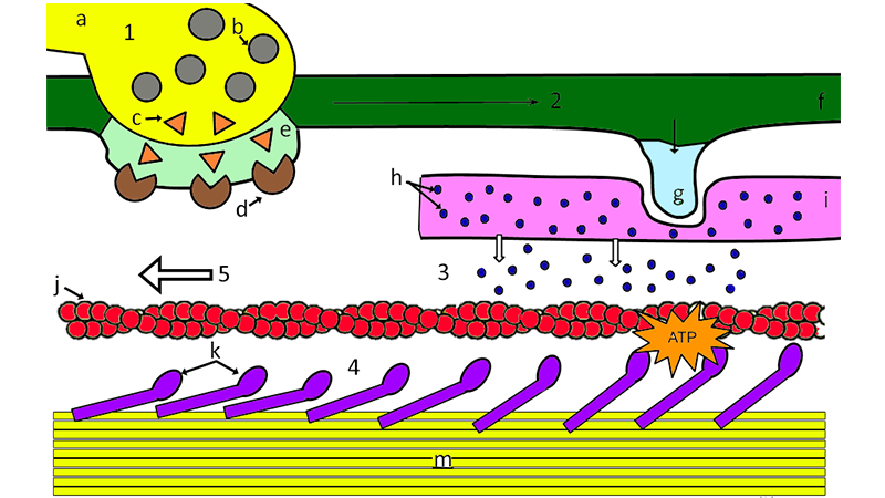
Sliding Filament Coloring
Step by step guide of the sliding filament model;, contraction starts with a nerve impulse and ends with muscle fibers shortening. Students color the diagram.
-
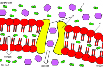
Cell Membrane Coloring
Color the cell membrane with a focus on diffusion, osmosis and transport proteins. Students color the structures of a cell membrane according to the directions. Then they answer questions about cell transport. I designed this worksheet for an introductory biology course to reinforce concepts related to cell transport. An image shows the phospholipid bilayer with…
-
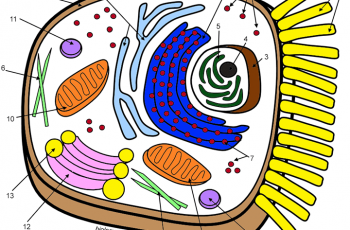
Learn the Animal Cell
This animal cell coloring worksheet can be used with freshman biology for years as a supplemental way to learn the parts of the cell. I assign it as a review or reinforcement exercise. It’s also a good activity for rainy days and sub days. This version of the cell coloring includes a cell diagram that…
-
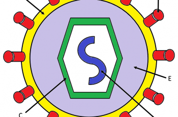
How Do Viruses Infect Cells (Coloring)?
A simple worksheet that explains how viruses infect cells which include diagrams to label and an image of a typical virus for students to color the envelope, proteins, DNA, and the capsid.
-

Photosynthesis Coloring
Students read short text passages and then color images to help them relate the textual information with the graphic.
-
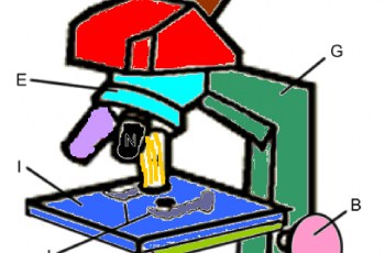
Color the Parts of a Microscope
Students read text that describe the parts and functions of the microscope and ask them to color the parts as they read.
-
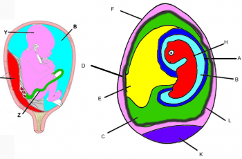
Comparing the Amniote Egg to the Placenta – Coloring
Color the amniote egg of a chicken and compare to the development of a human embryo.
-

Photosystems and Chemiosmosis Coloring
Use this coloring worksheet to explore how plant cells harvest energy form the sun to generate ATP in the process known as chemiosmosis.
-
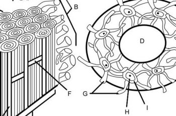
Bone Matrix Anatomy (Coloring)
Anatomy students learn about the skeletal system, where they examine bones and how the bones fit together to make up an entire skeleton. In addition, some course also explore bone tissue and how bone is formed, repaired, and even broken down to release minerals. The bone matrix is composed of cells called osteocytes…
-
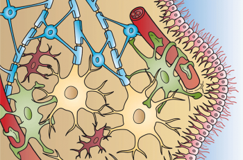
Color the Neuron and Neuroglia
Students can practice what they have learned about neurons with this simple coloring activity. The page shows features of the neuron, such as the axons and dendrites. They will also color the supporting cells of the matrix. There are no instructions, students must identify each of the types of glial cells: oligodendrocytes, astrocytes, microglial cells, ,…
-
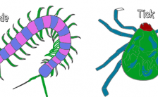
Comparing the Anatomy of Arthropods (Coloring)
This simple activity was designed for intro level biology (life science). Students read about different types of arthropods and learn what characteristics they share, such as an exoskeleton and segmentation. Then they reading focus on specific groups: insects, arachnids, crustaceans and centipedes. The goal is for them to learn that each group has a…
