Tag: label
-
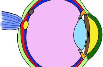
Anatomy of the Eye (Coloring)
The coloring worksheet is intended to help students learn the location of specific parts of the eye, like the cornea, sclera, lens, and retina.
-
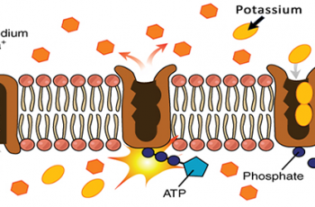
Cell Membrane Captions
Students examine images of transport across the cell membrane and identify key features such as the phospholipid bilayer, channel proteins, and receptors. Students then provide a title, such as “osmosis” and create a caption that describes the process being shown.
-
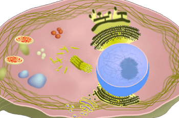
Cells Alive Worksheet
This worksheet follows diagrams and activities at CellsAlive.com which focuses on the size of cells compared to other objects, such as viruses and pollen. Students view interactive plant, animal, and bacteria cells to learn about the different structures associated with each.
-
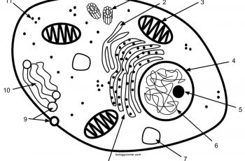
Cell Labeling: Simple and Complex
Students practice labeling organelles on a simple model (2D) and a more complex model. The idea is for students to gain an appreciation for how cell diagrams are created. They don’t all look alike, and are often artistically created. Cell organelles tend to follow basic design rules, like the mitochondria will generally look like a…
-
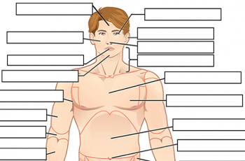
Label the Body Regions
This worksheet is used with a beginning anatomy unit that discusses anatomical terminology and body regions.
-
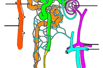
Color and Label the Nephron
Practice labeling the nephron with this reinforcement activity. Students can also color the image to identify the major structures of the nephron: glomerulus, bowman’s capsule, proximal and distal tubules, loop of Henle, collecting duct and capillaries. This was designed to go with a larger unit on how the urinary system and kidneys help the body…
-
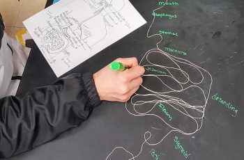
Modeling the Alimentary Canal
In this activity, students use string to model the gastrointestinal tract as a scale model. I’ve noticed that students do have difficulty with the concept of scaling, which is one of the crosscutting concepts listed in the NGSS. The directions give students measurements for a 1/3 scale model, the human alimentary canal is about 9…
-
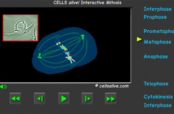
Mitosis – Internet Exploration
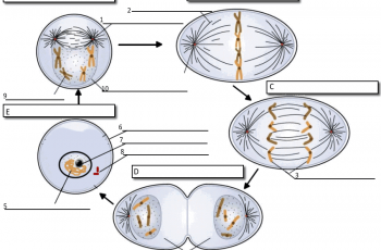
This assignment can be a stand-alone activity to help students learn to identify the phases of mitosis by viewing various animations. There are several sites to visit, where students perform tasks, such as labeling and making comparisons. Site 1: Bioman Mitosis Mover This is a game site where you progress through levels. Students can print…
-
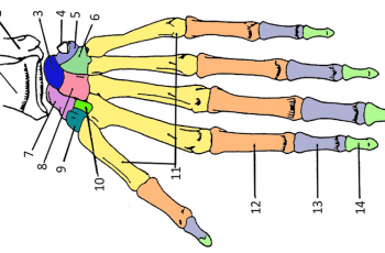
Explore Anatomy – Color the Bones of the Hand
Learning anatomy can feel daunting, but there are plenty of ways to make it more engaging and interactive. One great way to help students understand the structure of the human hand is through a hands-on (pun intended!) coloring exercise. By coloring the bones of the hand, students can reinforce their understanding of how these structures…
-
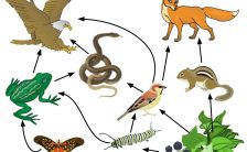
Food Web: Identify Consumers
Food webs are basic concepts in biology and ecology, where students learn the concept of energy flow in an ecosystem by viewing models of food webs. This labeling worksheet asks students to identify the primary, secondary, and tertiary consumers in a forest ecosystem. A food web is a representation of the complex interrelationship between…
-
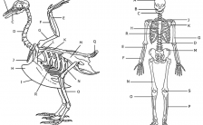
Comparing a Human and Avian Skeleton
Students often learn the bones of a human skeleton in health, but biology class can reinforce these lessons by comparing the human skeleton to that of other vertebrates. In this case, students color the skeleton of a bird and a human according to the directions. The colors will illustrate how many of the bones…
-
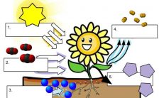
How Does Photosynthesis Work?
This handout can be used with a lecture on photosynthesis, where students label the main features of the light-dependent reaction and the Calvin cycle.
-
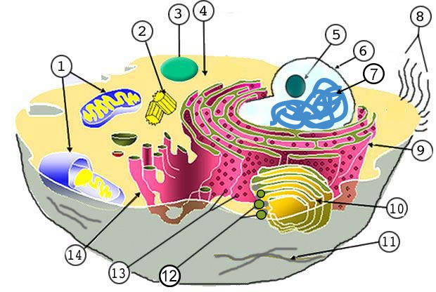
Label the Parts of the Plant and Animal Cell
Label a diagram of an animal cell and a plant cell; a diagram showing how proteins are produced by ribosomes, and finally packaged by the golgi apparatus.
-
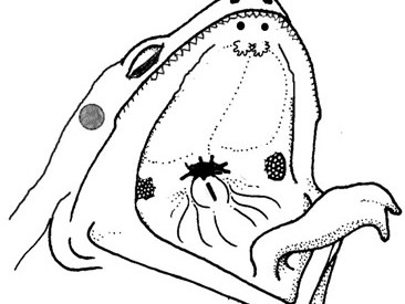
Student Guide to Frog External Anatomy
Lab handout over the external anatomy of the frog. Can be used as part of a frog dissection unit. Includes instructions and images to label.
