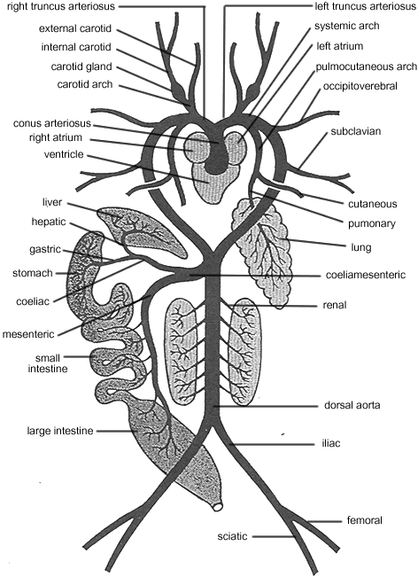
Circulatory System - Arteries


Where arteries and veins lie together, the arteries are generally more dorsal. In frogs, all main arteries emanate from the conus arteriosus, which divides into two arterial trunks inside the pericardial cavity. Each of these two trunks is generally called a truncus arteriosus.
Study and trace the left truncus arteriosus, carefully teasing away overlying muscles and membranes. The trunk soon divides into three main arteries: an anterior carotid arch, a middle, large systemic arch, and a dorsolateral pulmocutaneous arch.
Carotid Arch
The vessel branches a short distance after its origin into a smaller external carotid and a larger internal carotid. The ventral branch passes anteriorly along the floor of the mouth and into the tongue; it lies along side of the branches of the external jugular vein. The internal carotid posseses a prominent swelling at its base, called the carotid gland. The regulative structure aids in maintaining a sufficiently high pressure of blood flowing through the artery. This vessel leads into the brain.
Pulmocutaneous Arch
This dorsolateral vessel may be partly obscured by the large and more ventral systemic arch (in preserved specimens it may not be injected completely). Pull the left lung back and tease away overlying tissue to expose the pulmocutaneous more fully. Very near its origin, the vessel branches into two arteries; the pulmonary artery passing into the lung and the cutaneous artery passing laterally to the base of the foreleg - do not confuse it with the subclavian vein.
Systemic Arch
The truncus arteriosus becomes the systemic arch on both sides. It branches into the occipitovertebral artery, which supplies blood to the dorsal regions of the head, trunk and vertebral column. Just behind the occipitovertebral, the subclavian artery arises from the systemic arch. The subclavian supplies blood to the foreleg. The systemic arch then curves toward the midline where it joins the other systemic arch and becomes the dorsal aorta.
Dorsal Aorta
Just where the systemic arches unite into the dorsal aorta, the coeliamensenteric artery branches off. This vessel divides into the anterior coeliac artery and the more posterior mesenteric artery. Both pass through the mesenteries and supply parts of the alimentary canal. Thus the coeliac divides into the gastric, hepatic, and pancreatic arteries. The mesenteric divides into the splenic, anterior mesenteric, and posterior mesenteric arteries. Spend some time with these divisions until you locate them. Create a mental map in your head.
At the level of the kidneys, the dorsal aorta gives off short renal and genital arteries to the kidneys and reproductive organs. Along the lateral edge of the kidney cut through the peritoneum, which holds the kidney against the roof of the body cavity. Lift the kidney and note the arteries.
Posterior to the kidneys, the dorsal aorta divides into two iliac arteries to the legs. Trace these vessels out of the coelom and into the legs. Each iliac artery once it enters the leg soon divides into a sciatic artery and a femoral artery. The sciatic runs to the inside of the leg and the femoral runs to the outside of the leg.
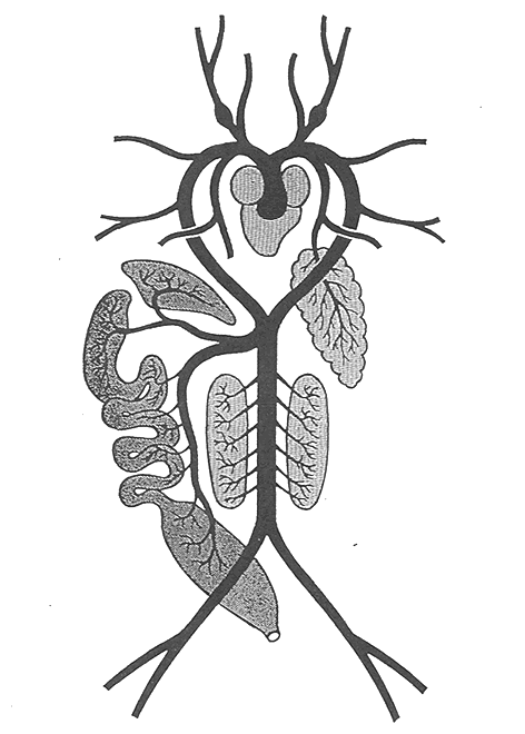
LABEL THE DRAWING
