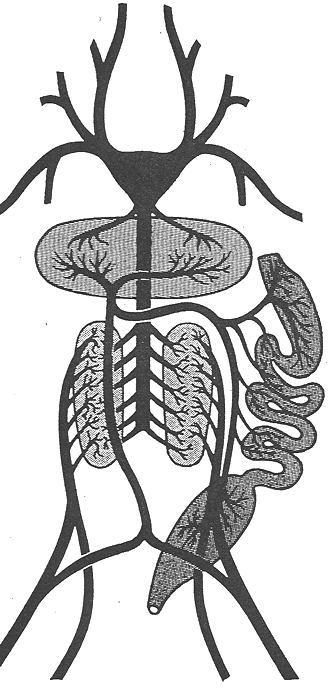
Dissection
of the Bullfrog Circulatory System - Veins

Observe the rules of good dissection even more closely than in the preceding sessions; you will be searching for very fine blood vessels and ducts that are difficult or impossible to locate unless you pay very strict attention to these directions.
Ventral Abdominal
Carefully trace this vein backward as far as possible, into the thighs. It is formed by the union of two pelvic veins come from the base of each thigh dorsally. Follow the vein forward and note its entry into the liver. In the liver, the vein breaks up into capillaries. If, at this point, the ventral abdominal vein has not been cut, you should cut it about 1 cm from where it enters the liver.
Hepatic Portal
Life the stomach forward and note that small veins from all parts of the intestine drain into a large vein which passes through the tissue of the pancreas. This is the hepatic portal vein. Trace it through the pancreas into the liver where it breaks up into branches to various lobes. In the liver lobes, these branches capillarize. (Note that veins beginning as capillaries and ending in capillaries are usually called portal veins. The hepatic portal begins in gut capillaries and ends in liver capillaries). The hepatic portal itself is formed by the union of the gastric vein (which goes to the underside of the stomach) and the mesenteric vein (which connects smaller vessels to the mesentery).
Hepatic
Cut transversely through the large intestine. This allows you to lift the major organs in locating various blood vessels. Lift the whole liver forward and near its attachment, try to locate the right and left hepatic veins that emanate from the liver and enter a very large vein near the posterior tip of the sinus venosus. Blood collected from the liver by the hepatic veins ultimately returns to the heart.
Renal Portal
Locate the left kidney, an elongated, reddish organ along the dorsal wall of the lower body cavity. On its ventral side lies a yellowish band of tissue, the adrenal gland, a component of the endocrine system. The adrenal gland may be impossible to locate in injected specimens (it just doesn't survive the process that well). Along the lateral edge of the kidney will be found the reasonably conspicuous renal portal vein. It enters the kidney and capillarizes there. The renal portal is one of the branches of the large femoral vein, the principal vein of the hind leg. Near the base of the leg, the femoral splits into the renal portal and a pelvis vein, which, as noted above, joins and becomes the ventral abdominal vein. The renal portal vein unites with a sciatic vein from the leg and passes to the kidney. Thus, follow the renal portal posteriorly until you find the juncture with the sciatic vein. The sciatic runs along the medial side of the leg and enters the body cavity near the cloaca. Posterior to this juction, find the union of the renal portal with the pelvic vein, and then locate the femoral vein in the leg.
Posterior Vena Cava
In the midline of the body between the two kidneys lies the largest of all veins, the posterior vena cava. Small, urogenital veins, sometimes called renal veins, enter it from the kidneys and the reproductive organs. Follow the posterior vena cava forward, for a short part of its course, it is embedded in the liver. Lift the liver forward and note the point of entry. Find also the point of exit, very near the sinus venosus. As noted, the hepatic veins from the liver join the posterior vena cava in this region.
At this point, if you wish, you may remove the posterior two-thirds of each lobe of the liver. Leave their points of attachment intact and do not damage the gall bladder.
Pulmonary
Relocate the left atrium and find the left pulmonary vein. It passes from the anterior edge of the left lung to the atrium. Just before its entry into the atrium, it joins the right pulmonary vein from the right lung. The pulmonary veins may not have filled with the blue latex in many specimens, but you may still be able to identify them
Anterior Vena Cava
The anterior vena cava and its associated veins also do not commonly fill with blue latex. You should make a serious effort to locate the blood vessels that are italicized in this section. You are responsible for being able to locate all vessels that are in italics on a drawing but not in an actual specimen.
Relocate the sinus venosus and not the large vein entering it laterally on each side. Each of these relatively short veins is an anterior vena cava. It is formed on each side by the union of three veins. The most posterior of these is the readily identifiable subclavian, which drains blood from the foreleg. Toward the anterior, lies the innominate vein, itself formed by the union of two branch veins near the angle of the jaw. The innominate vein splits into the subscapular (which drains the shoulder region) and the internal jugular. The external jugular is within the neck and drains blood from the mouth region and lower jaw.

