Bullfrog - External Anatomy


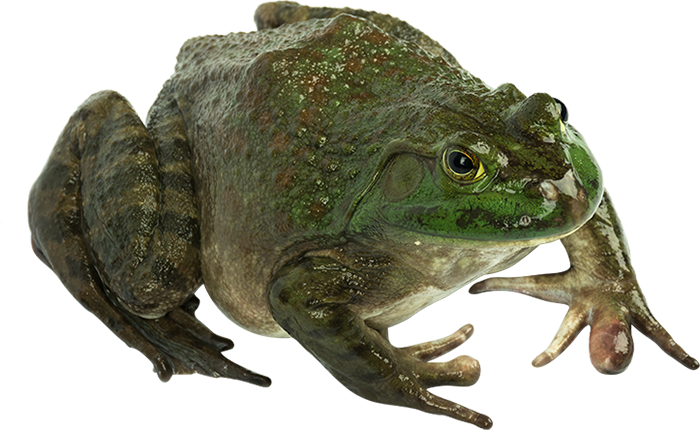
1. Trunk and Limbs
The principle body divisions are the head, trunk and limbs. Frogs are without neck or tail. The skin is smooth, moist in the living animal, and thin; is it also an important breathing organ. Slit the skin of the back and verify its thinness. Verify also the comparatively loose attachment of the skin to underlying body parts. In the living animal lymph fills the spaces underneath the skin. Note the coloration of the skin; pale ventrally and greenish with dark spots dorsally. To some extent a frog may change the depth of the greenish dorsal color according to the illumination of the background environment. The skin contains mucus secreting glands, concentrated particularly in the two dermal folds running longitudinally from behind each eye along the back to the base of the hind legs. Locate the anus at the posterior end of the trunk. Just in front and to each side of it along the back, pulsations of a pair of lymph vesicles may be observed in the living animal.
The front legs, as in man, consists of the upper arm, lower arm, wrist, hand, and fingers. During the breeding season (April - May), the inner finger of the male develops a black, swollen nuptial pad used in holding the female during mating. In the hind leg, identify the thigh, shank, ankle, foot, and webbed toes. Compare the digits of the hind leg with those of the foreleg.
Identify which thigh of the frog does NOT have a slit in it to allow for injection. Cut through the skin around the top of this thing and, using force, pull the skin from the leg in one entire piece. Examine the musculature; move the leg and note how the muscles change shape and length. With a probe, carefully loosen and separate individual thigh muscles from one another and locate the thigh bone, or femur. Along it lies the white sciatic nerve, the principal nerve of the leg, and the iliac artery, the principal blood supply to the leg. Trace the nerve and artery as far as the knee. In the shank, the largest muscle is the calf muscle, or gastrocnemius. Loosen it away from the other muscles and note its origin at the knee and its insertion on the foot. Insertion is achieved by the prominent Achille's tendon, which passes behind the heel and spreads fan-like over the sole of the foot. Pull on the gastrocenemius and observe the resulting motion of the foot.
2. Head, Mouth and Pharynx
The head bears three pairs of prominent sense organs, namely, eyes, nose, and ears. The bulging eyes are equipped with a more or less non-movable upper lid, and a non-moveable lower lid, and a transparent fold, the nictitating membrane. The latter is attached to the inside of the lower lid and can be moved up over the eye. A frog can close its eyes altogether by retracting them deep into their sockets, a process which also brings the upper and lower lids together. Ear the tip of the snout are the nostrils, or external nares. Pass a probe through the nostril to locate the internal nares found at the roof of the mouth. Behind each eye locate the circular eardrum, or tympanum; outer ears are not present in frogs. In the center of the tympanum a slightly raised spot marks the attachment of the interior columella, a single middle ear bone transmitting sound waves to the inner ear (there are 3 middle ear bones in the mammal).
Put the frog ventral side up in your dissecting pan and pin down hands, feet, and upper jaw. With bone shears, cut through the angles of the jaws on each side so that the lower jaw can be folded back to expose the mouth cavity. On the floor of this cavity is the tongue, attached at its anterior end to the lower jaw: a frog hurls its sticky tongue out hind end first. Along the rim of the upper jaw feel with your finger for the teeth: premaxillary teeth along the middle, and maxillary teeth along the sides. Another set, called vomerine teeth, is present on the roof of the mouth and is attached to skull bones called vomers.
Note the pharynx, the space behind the mouth cavity toward the throat. Depress the floor of the mouth and probe carefully into the pharynx until you find a median projection with a longitudinal slit. The projection marks the position of the larynx (voice box) and the slit is the glottis, through which air passes into and out of the larynx - and from there directly to the lungs. Probe between the larynx and the roof of the pharynx along the midline of the body and find the opening into the esophagus, the alimentary tube leading to the stomach. In the angles of the jaws, on a level with the esophageal opening, locate the opening of the Eustachian tube on each side. These tubes connect to the middle ear and help to equalize pressure when the frog dives in water. Verify the connection between the ear and the Eustachian tubes by slitting open the tympanum. Again, note the columella. Pass a probe through the ear canal to see where it leads.
Make a drawing
of the frog's open mouth and label all appropriate structures. You may wish
to consult supplementary materials for your drawing.
