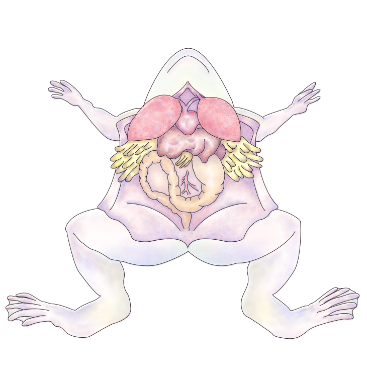
Dissection of the Bullfrog - Internal Anatomy

The Coelom
Close the mouth of the frog and pin both jaws down. Life the ventral trunk skin with forceps or needle, and with scissors make a longitudinal slit through the skin along the midline. Extend this slit forward into the lower jaw and backward to the end of the trunk. Make lateral cuts to each side, one set in front of the forelegs and another near the base of the hind legs, and pin the resulting flaps to the side. Note the ventral body-wall muscles, and along the mid ventral line, the ventral abdominal vein.
Lift the muscles near the posterior end of the trunk and make a longitudinal incision through the muscles slightly to the left side (your right) of the vein. Then (always lifting the parts to be cut to prevent damage to underlying structures) continue the incision forward to the level of the forelegs, where you will encounter the transversely placed bones of the pectoral girdle.
Raise the girdle with forceps (do not be afraid to exert force) and continue your incision by cutting through the sternum (breastbone). Now make lateral cuts through the body wall to each side, one set of cuts immediately behind the pectoral girdle, another set at the posterior end of the trunk incision. Fold to the side and pin down the resulting flaps of body wall muscles. Also fold to the side the left and right portions of the cut pectoral girdle.
The coelom, or body cavity, and most of the internal organs are now exposed. Note the shiny peritoneum lining the inner surface of the body wall muscles. This thin membrane also covers and holds in place all other internal organs. Those peritoneal portions that fold around and hold such organs are called mesenteries.
If your specimen is female, much of the coelomic cavity is likely to be filled by a pair of large, transparent ovaries, each containing hundreds of black and white eggs. If the ovaries are full of eggs, gently lift the left overy with forceps and finds its place of attachment. Cut through the attachment and remove the ovary in one piece. If the other ovary still obstructs your view, it too may be removed.
In freshly killed specimens, one or both lungs may be highly inflated with air and appear as elongated, reddish sacs. Since the lungs are not usually inflated in preserved specimens, they will be partly hidden under the liver, the large, prominent, dark brown organ in the midventral portion of the trunk.
The Heart
If necessary, lift or shift the liver gently until you locate the heart. On each side of the heart is a lung. The heart lies in a special part of the coelom, called the pericardial cavity. This space is separated from the remainder of the coelom by a membranous pericardium. Lift the pericardial membrane with forceps to slit it open and expose the heart fully. ( Due to the injection process, this membrane may be partially destroyed. )
Identify the single thick-walled, triangular ventricle and the two more anterior, thin walled atria. Lift the apex of the heart and snip the slender strand of tissue that connects the atria to the dorsal wall of the pericardium. In this dorsal atrial region, well underneath the ventricle, locate the sinus venosus, a thin walled, membranous sac which collects all blood coming to the heart from the veins of the body.
Three large veins enter the sinus venosus: the posterior vena cava (along the midline), and the anterior vena cava (laterally on each side). The sinus venosus itself empties into the right atrium (even though it appears to connect to both atria).
The left atrium receives blood returning from the lungs via the pulmonary veins. These lie dorsal to the sinus venosus and will be difficult to see at this stage. Let the heart fall back to its normal position and locate the conus arteriosus, a single wide arterial vessel emanating from the ventricle and passing ventrally over the right atrium. Follow it forward to where it divides into two branches, each known as the truncus arteriosus.
Make labeled drawings of the frog's heart (or label the ones provided) one dorsal view and one ventral view. Again, you may wish to consult supplementary material.

