Dissection
of the Bullfrog

Introduction

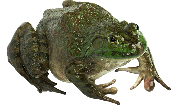
In this laboratory exercise, the anatomy of the bullfrog will be examined in some detail. You may recall that in your first year biology course you dissected a very similar organism knows as the grass or leopard frog (Rana pipiens). The bullfrog (Rana catesbeiana) which we will be using is much larger than the grass frog, enabling us to find and relate to more structures.
The classification of the bullfrog
Kingdom Animalia
Phylum Chordata
Subphylum Vertebrata
Class Amphibia
Order Anura
Family Ranidae
Genus Rana
Species catesbeiana
The lab books and
diagrams available to you are supplemental. You are expected to follow the directions
in this lab. You will be held responsible for being able
to locate all the underlined structures in this handout. Several other supplemental
texts are available; feel free to ask to use them. You are expected to have
exhausted all possibilities in attempting to located structures before asking
for assistance. Using the available material, instructions and diagrams, most
students will be able to locate most structures for themselves. If after an
earnest effort, you cannot find a structure, ask for assistance. Remember, this
is a learning experience, it is quite permissible to discuss and observe other
students' specimens. Compare you dissection with others, for animals often differ.
Make sure to look at one of the opposite sex. In addition to the external anatomy
of the frog, we will emphasize three systems.
- Digestive System
- Circulatory System
- Urogenital System
The specimen you will receive is a preserved double-injected specimen. Double injected refers to the arteries being filled with a red latex, and the veins being filled with blue latex. You will notice various incisions on the external surface of the frog where the latex was injected.
The frog is a vertebrate, which means that many aspects of its structural organization are common with all other vertebrates, including man. The similarity of structures among related organisms shows evidence of common ancestry. In a way, studying the frog is like studying a human. As the leading theme of this lab, ask yourself: for every structure observed in the frog, there is an equivalent structure in your own body - what is the structure and where is it located.
As the second leading theme, pay particular attention to the relationships among organs and groups of organs. Structural parts are not "just there" in random locations. Their specific layout within the body contributes to making certain functions possible. Therefore, for every structure seen, you should determine the following:
- What organ system it belongs to
- How it is connected with other components
- Its general function
- Its specific function (if applicable)
Dissection
Dissecting tools will be used to open the body cavity of the frog and observe the structures. Keep in mind that dissecting does not mean "to cut up"; in fact, it means "to expose to view". Careful dissecting techniques will be needed to observe all the structures and their connections to other structures. You will not need to use a scalpel. Contrary to popular belief, a scalpel is not the best tool for dissection. Scissors serve better because the point of the scissors can be pointed upwards to prevent damaging organs underneath. Always raise structures to be cut with your forceps before cutting, so that you can see exactly what is underneath and where the incision should be made. Never cut more than is absolutely necessary to expose a part.
Grading
Your grade on this laboratory will be assessed according to the following criteria
- Class Participation (observed daily)
- Quizzes throughout
- Lab Practical Exam (at the end of lab)
- Dissection of the Bullfrog Lab Questions
Glossary of Terms
Dorsal: toward
the back
Ventral: toward the belly
Lateral: toward the sides
Median: near the middle
Anterior: toward the head
Posterior: toward the hind end (tail)
Superficial: on or near the surface
Deep: some distance below the surface
Sagittal: relating to the midplane with bisects the left and right sides
Transverse: relating to the plane separating anterior and posterior
Horizontal: relating to the plane separating dorsal and ventral
Proximal: near to the point of reference
Distal: far from the point of reference
Pectoral: relating to the chest and shoulder region
Pelvic: relating to the hip region
Dermal - relating
to the skin
Longitudinal - lengthwise

