Dissection of the Bullfrog - The Urogenital System


The excretory and the reproductive system of the frog are combined into a system called the urogenital system because so many of the excretory and reproductive structures and paths are shared. You will be responsible for knowing the structures for both a male and female frog. When you are finished with the dissection of your frog, trade with another group in the room to view a frog of the opposite sex.
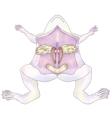
Female
Examine the remaining ovary and find its attachment to the mesenteries to the medial border of the kidney on that side. Attached to each ovary anteriorly is a fat body, a yellowish bundle of finger-like strands. This fat storing organ varies in size seasonally, being largest in the summer. Underneath each ovary will be found the thick, whitish, highly coiled oviduct. Trace the left oviduct forward to its beginning, well under and near the base of the lung. The anterior oviduct opening is the funnel-like ciliated ostium. Ripe eggs are released from the ovaries into the body cavity at one time during breeding season, the eggs then enter and pass through the oviducts. Near the posterior border of the kidneys, each oviduct widens into a thin-walled ovisac, where descending eggs may be stored temporarily. Carefully separate the left half of the urinary bladder from its attachment to the left ovisac and examine the shape and extent of this sac. Posteriorly, it joins the right sac along the midline.
Along the edge of the kidney, find a thin white tube. This is the ureter which carries urine from the kidney. Trace the duct forward along the kidney as far as you can see it and backward to its termination in the cloaca. The urinary opening in the cloaca itself is usually too small to be seen.
Male
At the end of each kidney (toward the anterior) lies a testis, a rounded yellowish organ. Attached to it is a fat body, a yellowish bundle with finger-like strands. This fat storing organ varies in size seasonally, being largest during the summer. Closely examine the dorsal surface of the mesentery that holds the testis to the kidney. Note the fine blood vessels and between them, minute white ducts passing from the testis to the kidney. These are sperm ducts, or vasa efferentia. They conduct sperm into kidney tubules that also carry urine. Along the lateral edge of each kidney find a prominent wavy tube, the muellerian duct. This is functionless in the male and is equivalent to the oviduct found in the female - it is also called a vestigial oviduct. Locate the tube leading from the kidney to the bladder, this tube carries urine and is called the ureter. At the base of the ureter, you'll find an enlarged area, the seminal vesicle; it is used for storing sperm. Sperm eventually exit out the cloaca.
