Investigation: Exploring Different Types of Cells


All living organisms are made of cells. The smallest cells are about 0.001 millimeters in diameter and belong to one of two domains: Bacteria and Archaea. Organisms in these groups do not have a nucleus or membrane-bound organelles. These organisms were some of the first to live on the planet; archaea lived over 3.5 billion years ago.
Organisms can belong to a third domain: Eukarya. Eukaryotic cells are larger (0.01 to 1 mm) and contain a nucleus and organelles, such as mitochondria and endoplasmic reticulum. Plants, animals, fungi and protists belong to this domain.
In this investigation, you will view both preserved specimens and living specimens from the domain Eukarya and the domain Bacteria.
Exercise 1: Getting to Know the Domain Bacteria
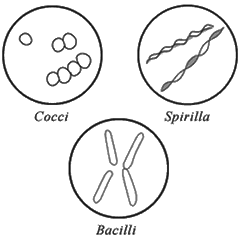
Bacteria cells are very small compared to eukaryotic cells. They are visible with a light microscope using the high power objective. Bacteria come in four shapes: cocci (round), spiral, and bacillus (rod-shaped). The are often named based on their shape. For example, round bacteria that occur in chains are called “Streptococcus” and if they occur in clumps, they are called “Staphylococcus.” E. coli , which is a bacillus, is about 2.0 µm long and .5 µm wide. .
View prepared slides of bacteria at 400x. Sketch the bacteria to scale and describe their appearances. Pay attention to detail with regard to the size of the cell and their shape and coloration.
Specimen (name on slide) |
|
|
Sketch (400x) |
|
|
Description |
|
|
Exercise 2: Getting to Know the Domain Eukarya
1. Create a wet mount of your own cheek cells. (Kingdom Animalia)
 Add a small drop of methylene blue to a clean slide.
Gently scrape the inside of your cheek with a toothpick and then smear the sample in the dye.
Place a cover slip over the dye/sample and view.
Cells will appear as small light blue with a darker area in the center (nucleus)
They will appear very small at 40x.
When finished, throw the entire slide away.
Sketch the cheek cells at each of the magnifications (to scale) and label the nucleus, plasma membrane and cytoplasm of the 400x sample (high power)
Estimate the diameter of a cheek cell: _______________________
3. Create a Wet Mount of Onion Cells (Kingdom Plantae)
Cut an onion and remove the thin skin-like membrane that lines the onion layers with tweezers.
Place a tiny portion of this skin a slide and stain with iodine.
Place a cover slip over the sample and view at 40x, 100x and 400x.
Sketch the onion cells and label the cell wall and the nucleus of the sample at 400x.
Estimate the length of a single onion cell. ___________
4. View Plant Cell Leaves (Kingdom Plantae)
Prepare a wet mount of an elodea leaf by placing a single leaf on a slide with a drop of water.
Place a cover slip over the sample and view at 400x..
You will notice many green blobs within your cells, these are chloroplasts. The nucleus will appear brownish-red in color, but it may be obscured by the many chloroplasts within the cell.
You may also notice that the chloroplasts move around a large central vacuole. This movement is a result of cytoplasmic streaming.
Sketch the elodea cells at 400x and label the cell wall and the chloroplasts of the sample at 400x.
Estimate the length of an elodea cell: _____________
5. View Protozoa (Kingdom Protista)
a) There are many preserved slides of protozoans available which include the ameba, paramecium, euglena and spirogyra. You can choose to focus on one or two of these samples.
b) Sketch and label the protozoan(s) you chose to investigate.
c) Choose one of your samples and estimate its size. ____________
Specimen (name on slide) |
|
|
Sketch (400x) |
|
|
Description |
|
|
Exercise 3: Application
1. Compare and contrast prokaryotic cells and eukaryotic cells, listing at least 3 similarities and 3 differences.
2. Compare and contrast plant cells and animal cells, listing at least 3 similarities and 3 differences.
3. You viewed two plant specimens (onion and elodea leaf). How did these cells differ and why might organelles like chloroplasts be present in one, but not the other. (Hint: think about where these plant structures grow in relation to the overall plant.)
3. Much of this investigation asked you to estimate the sizes of cells you viewed. Create a list of cells, ranked in order from the smallest to the largest that includes at least 4 of the cells you observed in this investigation.
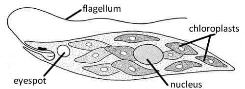
4. The Kingdom Protista traditionally contains microorganisms that are difficult to classify as plants or animals. In fact, one protist, called the euglena can switch from being a heterotroph to being an autotroph depending on food availability. The image below shows a euglena. Discuss why biologists might have a difficult time classifying this organism.
Other Laboratory Activies on the Cell
Onion Root Tip Lab – view real cells with a microscope, requires lab equipment and prepared slides
Cheek Cell Lab – observe cheek cells under the microscope
Comparing Plant and Animal Cells – compare onion cells to human cheek cells
Why Are Cells So Small? – measure the surface area and volume of boxes as a cell model
Protozoan Slide Set (from Amazon)