Simple Worms - Microscope Observations
 Vinegar Eel
-- When you get your slide, smell the drop of water on it. This should give you a clue as to how these nematodes got their name. Nematodes that are commonly referred to as "vinegar eels" (Turbatrix aceti) exist by feeding on bacteria and fungi found in the sediments of nonpasteurized vinegar. When viewed alive, vinegar eels are seen to be in constant motion.
1. Where do vinegar eels live?
2. How many nematodes do you think are on your slide?
3. Sketch the shape of the nematode. Is there an obvious anterior side (or head)?
Nematoda (roundworms)
Trichinella Worm -- slides will show infected muscle tissue
Trichinellosis, also called trichinosis, is caused by eating raw or undercooked meat of animals infected with the larvae of a species of worm called Trichinella. Infection occurs commonly in certain wild carnivorous (meat-eating) animals but may also occur in domestic pigs. Trichinella worms belong to the Kingdom Animalia and the Phylum Nematoda.
When a human or animal eats meat that contains infective Trichinella cysts, the acid in the stomach dissolves the hard covering of the cyst and releases the worms which mature and eventually make their way back to the muscles, the worms curl into a ball and encyst (become enclosed in a capsule).
Obtain a slide of muscle tissue infected with trichina cysts. View the slide under scanning and low power. Look for long striped muscles with circular cysts embedded within.
4. Sketch the muscle tissue and use an arrow to indicate where the trichina cysts are located.
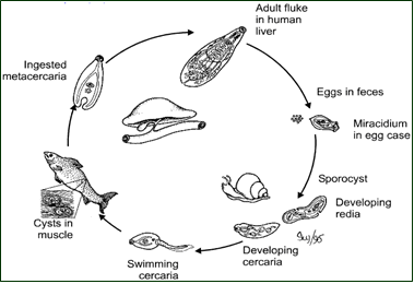
Schistosome Worm - obtain a slide of the schistosome worm, but note that these worms (flukes) have several forms depending on the stage of their life cycle.
5. Sketch your worm
6. Pictured is a diagram of the liver fluke. Two intermediate hosts (vectors) are shown; what are they?
Observe the Tapeworm - obtain a slide of the tapeworm and view under the microscope.
7. To what phylum
does the tapeworm belong?
Kingdom?
8. Sketch the tapeworm. Label the proglottids, the scolex, and the hooks.
Related Resources
Comparing Plant and Animal Cells – compare onion cells to human cheek cells
Microscope Investigation for AP Biology – refresher on how to use a microscope and how to measure viewing field
Exploring Cells – follow in the footsteps of famous scientists like Hooke and Van Leeuwenhoek by looking at slides of cork, paramecium (animalcules) and typical plant and animal specimens.
Investigation: Banana Starch and Plastids – compare a ripe banana to a green banana by viewing plastids stained with iodine
Rotifer Lab - observe living rotifers with the microscope