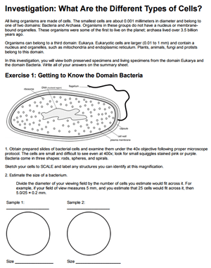
This lab requires students to look at cells from different domains and kingdoms. Students compare the size of cells within the eubacteria domain to the size of eukaryotic cells from the plant and animal kingdoms. Ideally, students will have already completed “Investigation: Microscope” so that they are familiar with making scientific sketches and estimating sizes.
This investigation provides a survey of the many basic types of cells the students in biology may encounter. They also compare the cells found in the roots of plants, to cells found in the leaves.
I use prepared slides of bacteria and protozoans. Bacteria can be difficult to view due to their small size. To make this part of the exercise more engaging, I like to provide students with pathogenic bacteria. This can lead to engaging discussions about botulism and tuberculosis!
Protozoans are usually fun for students to view because they come in so many different shapes and sizes. You can order prepared slides or even cultures with living specimens. Paramecium and ameba are my favorites!
Students then create their own slides of their cheek cells and onion cells. Students should already know how to stain cells from an earlier microscope lab. I use methylene blue for cheek cells and iodine for onion.
Instructors can modify this lesson dependent upon what resources they have available.
I encourage students to photograph their slides using their cell phones and upload them to Google classroom. We can then view them as a class and have a discussion about similarities and differences.
Grade Level: 9-12 | Time: 2-3 hrs
HS-LS1-2 Develop and use a model to illustrate the hierarchical organization of interacting systems that provide specific functions within multicellular organisms

