Microscope Activities

Introduction to the Microscope – E Lab – explore how to use a light microscope (Letter E Slides)
Microscope Coloring – label and color the parts of the microscope
Label the Microscope – simple graphic to practice identifying microscope parts
Guide to Using the Light Microscope – step-by-step instructions on how to focus images and create wet mount slides.
Microscope Investigation for AP Biology – refresher on how to use a microscope and how to measure viewing field
Cheek Cell Lab – observe cheek cells under the microscope
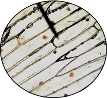
Observing Plant Cells – microscope observation of onion and elodea
Comparing Plant and Animal Cells – compare onion cells to human cheek cells
Investigation: What are the Different Types of Cells – survey of different kinds of cells, cheek, blood, onion root, leaf
Exploring Cells – follow in the footsteps of famous scientists like Hooke and Van Leeuwenhoek by looking at slides of cork, paramecium (animalcules) and typical plant and animal specimens.
Investigation: Banana Starch and Plastids – compare a ripe banana to a green banana by viewing plastids stained with iodine
Exploring Eukaryote and Prokaryote Cells – a comprehensive microscope lab that covers various types of cells like bacteria, plants, animals, and protozoa.
Why Are Cells So Small? – measure the surface area and volume of boxes as a cell model
Cell Size Experiment – use agar and a basic solution to illustrate how fast fluids can diffuse into a cell, dependent on cell size
Comparing Plant and Animal Cells – how are they alike, and how are they different?
Coloring and Labeling Activities

Animal Cell Coloring – color a typical animal cell
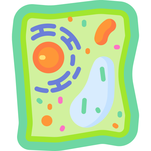
Plant Cell Coloring – color a typical plant cell
Prokaryote Coloring – color a typical bacteria cell
Cell City Analogy – compares a cell to a city by describing what each part of the cell does
Internet Activity on Cells (Cells Alive) – view interactive cells at cellsalive.com; complete worksheet
Cell Model – create a cell from household and kitchen items, rubric included
Cell Rap – fun poem to describe the parts of the cell, sing to the tune of “Fresh Prince”
Practice Labeling the Cell and Endomembrane System – complex drawing showing plant and animal cells, and protein synthesis
Simple Cell Labeling – simple cell drawing compared to a 3d complex cell diagram
Amoeba Sisters Video – worksheet for short program that outlines the structures of the cell and their functions
Amoeba Coloring – a single celled protist that moves using pseudopodia
Paramecium Coloring – a single celled protist that moves using cilia
Euglena Coloring – a single celled protist that moves using flagella, and can be both autotrophic and heterotrophic
 Cell Readings and Data Analysis
Gaucher Disease – explores a lysosome storage disorder
Mitochondria and Energy Production – explores structure of the mitochondria and how ATP is harvested
Data Analysis of Mitochondria Function – explores oxidative damage resulting from a toxic exposure (AP Bio)
What Causes Mitochondrial Disease – CER that explores different types of mutations that results in nonfunctioning mitochondria
Mitochondrial Disorders – What Happens When Cells Don’t Get Enough Energy – read about a young girl diagnosed with a mitochondrial disorder (causes and symptoms)
Infectious Disease Project – design an infographic exploring an infectious disease
 Cell Reproduction
Mitosis in an Onion – view picture, identify the stages of mitosis in each of the cells
Cell Cycle Label – label a picture of the stages of mitosis, identify parts of the cell such as the centriole and spindle
Onion Root Tip Lab – view real cells with a microscope, requires lab equipment and prepared slides
Onion and Whitefish – view cells, if you missed the classroom lab; virtual version of the mitosis lab
Mitosis Internet Lesson – view animations of mitosis; questions
Meiosis Study Guide – a list of questions to consider when studying meiosis
Reinforcement: Meiosis – focus on vocabulary, such as gamete and zygote, diploid and haploid; label an image
Meiosis Internet Lesson -view animations and tutorials, with questions
Identify the Phases of Meiosis – descriptions of what occurs in each phase, label an image
Phases of Meiosis – describe what is happening in each phase (chart) and label the phases
Cell Cycle Cut and Paste – students arrange words and draw arrows to illustrate mitosis
Cancer: Out of Control Cells – article describing how the cell cycle relates to cancer, includes questions
Osmosis and Diffusion
Cell Membrane and Transport – student notes and Google slides (Bio 1)
Observe Diffusion in a Bag – model diffusion using a plastic baggie, iodine and a beaker. This article explains what happens.
Story Lab – How Can Diffusion be Observed – based on the “diffusion in a bag” lab where students read a passage about diffusion
Investigation: The Effect of Salt on a Potato – simple lab where a potato is soaked in salt water.
Cell Membrane and Transport Coloring – shows the phospholipid bilayer and different types of transport
Transport Across the Cell Membrane – simple diagram shows how molecules enter the cell through diffusion, facilitated diffusion, and osmosis
Cell Membrane Images – work in groups to create captions and titles for images depicting the cell membrane and transport across it.
Investigation: Why Are Cells So Small? – use bromothymol blue and agar to model how diffusion occurs in cells
Case Study: Cystic Fibrosis – for AP Biology, examines the role of cell membrane proteins in clearing mucus from the lungs.
Observing Osmosis – use an egg, vinegar, corn syrup, will take a few days
Salt and Elodea – quick lab to observe the effects of salt water on elodea cells
Diffusion and Osmosis Crossword Puzzle – vocabulary review
Cell Transport Review Guide – with questions, definitions and images
Osmosis in Cells – AP Lab 1, modified, using dialysis tubes and sugar solutions to observe water movement
Water Potential and Molarity – measure the mass of carrots after soaking in different molarity solutions to determine the molarity of the carrot
Investigation – Cell Membrane Bubble Lab – model properties of the cell membrane with straws and a bubble solution
Cellular Respiration
Photosynthesis and Respiration Model – image shows the two processes are connected; students answer questions related to a graphic
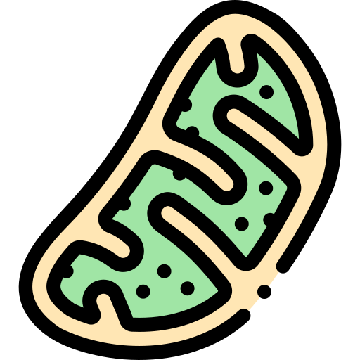
Color the Mitochondrion – guides to the process of cellular respiration, with coloring!
Chemiosmosis Coloring – color the membrane and the steps involved in the production of ATP
Converting Light Energy into Chemical Energy – label of graphic of photosystems I and II
Cellular Respiration Graphic Organizer – map of the steps involved in cellular respiration (Key, TpT)
Cellular Respiration Data Analysis – interpret graphs showing rats of respiration
Observing Cellular Respiration – AP Biology Lab using peas and respirometers
Cellular Respiration Virtual Lab – AP Lab that can be performed online at LabBench
Rate of Respiration Virtual Lab – use a simulator to show change in respiration of germinating seeds
Case Study: The Cyanide Murders – explores cellular respiration and why we need oxygen
Mouse Respiration CER – shows a graph of respiration rates at different temperatures
Photosynthesis
Plant Pigments – Use chromatography to observe plant pigments separate
Photosynthesis Simulation – this simulator uses light and varying levels of carbon dioxide to explore rates of photosynthesis, replaces the waterweed simulator
Photosynthesis Lab – AP Lab, uses spinach leaves and light to measure the rate of photosynthesis. Oxygen bubbles cause leaf disks to float when they are exposed to light.
Calvin Cycle (TED-Ed) – video worksheet with labeling and practice questions
Inquiry Investigation – what factors affect rate of transpiration – use leaves and evaporation to explore how plants take up water
Stomate and Leaf Investigation – view stomata under the microscope and determine density on upper and lower surface of the leaf
Investigate Photosynthesis with Vernier Probes – explore changes in oxygen levels in plants exposed to light
Use Phenol Red to Observe Photosynthesis – submerge water plants in a solution of phenol red, color change indicates changes in carbon dioxide in solution
Cell Metabolism and Biochemistry
Acids and Bases – basic coloring showing how water dissociates into ions, pH scale
Biomolecules Boxing – students make boxes showing the four biomolecules of life and use the boxes to practice structure and functions
Enzyme Lab – use liver to show how catalase breaks down hydrogen peroxide into oxygen and water, bubbling is used to measure the reaction at different temperatures.
Elements Found in Living Things – a simple coloring exercise that shows the percentage of the elements in organisms (CHNOPS)
Analyzing Graphics – Enzymes – shows substrates and enzyme interactions and explores competitive inhibition
Observe Catalase Activity in Yeast – create sodium alginate spheres to observe how catalase breaks down hydrogen peroxide
Enzyme Activity Using Toothpickase – simulate the activity of an enzyme by breaking toothpicks.
Organic Compounds – concept map, students fill in terms related to organic chemistry (carbohydrates, nucleotides..etc

